
The heart sits inside a double-layered protective sac called the pericardium. Under normal conditions, a small amount of fluid — typically between 15 and 50 milliliters — fills the space between those two layers, lubricating the heart as it beats and reducing friction against surrounding tissue. When that fluid accumulates beyond normal levels, the condition is called a pericardial effusion.
The amount of fluid matters enormously, but so does the rate at which it accumulates. A large effusion that develops slowly over weeks or months may cause little noticeable discomfort because the pericardium has time to stretch. A smaller effusion that develops rapidly — from trauma or a sudden infection, for example — can compress the heart severely and become life-threatening in hours. This is why pericardial effusion is a condition that ranges from an incidental finding on a routine chest scan to a cardiac emergency requiring immediate intervention.
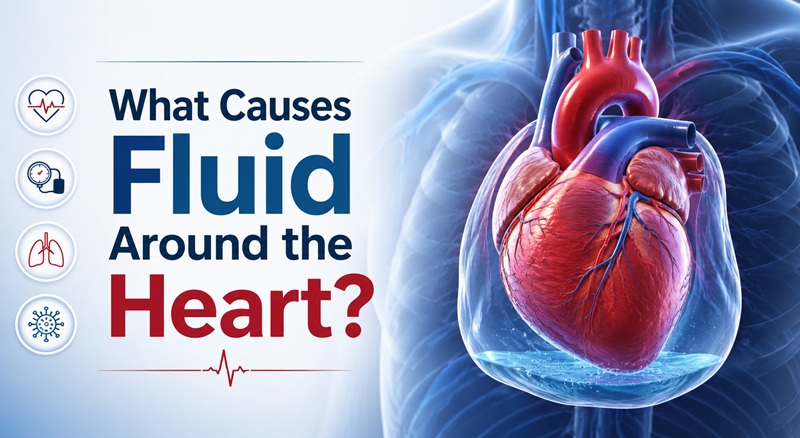
Understanding what causes fluid to collect around the heart is the first step toward understanding when it is dangerous, how it is diagnosed, and how it is treated.
The Anatomy Behind the Problem
The pericardium is not a single layer but a two-layer structure: an outer fibrous layer that anchors the heart in the chest and an inner serous layer that itself consists of two membranes with a small fluid-filled cavity between them. This cavity is the pericardial space, and the fluid within it is pericardial fluid.
This arrangement serves the heart well under normal conditions. Problems begin when something disrupts the balance between how much fluid enters the pericardial space and how much is reabsorbed. That disruption can come from inflammation that causes the pericardial membranes to leak, from cancer cells that invade the pericardium and block fluid drainage, from bleeding into the pericardial space after trauma or surgery, from systemic diseases that cause fluid to accumulate throughout the body, or from kidney or thyroid conditions that alter fluid balance and protein levels.
Each of these mechanisms produces fluid with different characteristics — inflammatory effusions tend to be protein-rich and cellular, while effusions from heart failure or low protein states tend to be thin and watery. The type of fluid helps physicians identify the underlying cause.
The Most Common Causes
Idiopathic — Unknown Cause
In developed countries, the most frequently diagnosed cause of pericardial effusion is idiopathic — meaning no specific cause is identified despite investigation. This category accounts for a substantial proportion of cases, and most of them are thought to represent viral pericarditis that was never diagnosed as such because the viral illness was mild, brief, or attributed to something else. When a patient presents with a small-to-moderate effusion and no obvious underlying disease, a viral cause is often assumed even without laboratory confirmation.
Idiopathic effusions are generally managed conservatively with anti-inflammatory medications and tend to resolve without recurrence in most patients.
Infections
Viral infections are the most common infectious cause, with viruses such as Coxsackievirus, Echovirus, Epstein-Barr virus, cytomegalovirus, influenza, and SARS-CoV-2 among those known to trigger pericardial inflammation and fluid accumulation. The mechanism is immune-mediated inflammation — the immune response to the infection irritates the pericardium and causes it to leak fluid into the pericardial space.
Bacterial infections can also cause pericardial effusions, and these tend to be more serious than viral causes. Tuberculosis is one of the leading causes of pericardial effusion globally, particularly in regions where TB remains prevalent. TB-related pericardial effusion can be large, difficult to treat, and carries a risk of long-term complications including constrictive pericarditis, in which the pericardium becomes scarred and rigid, impairing cardiac filling.
Fungal infections, while less common, can cause pericardial effusions in immunocompromised patients, including those on long-term steroids, receiving chemotherapy, or living with HIV/AIDS.
Inflammatory and Autoimmune Conditions
Diseases in which the immune system attacks the body’s own tissues are a major cause of pericardial effusion. Lupus (systemic lupus erythematosus) is the autoimmune condition most frequently associated with pericardial involvement — pericarditis and effusion occur in approximately 25 percent of lupus patients at some point in their illness.
Rheumatoid arthritis, Sjögren’s syndrome, inflammatory bowel disease, sarcoidosis, and other systemic inflammatory conditions can all cause pericardial inflammation and secondary fluid accumulation. In these cases, treating the underlying autoimmune condition is central to resolving and preventing recurrence of the effusion.
Dressler syndrome — also called post-myocardial infarction syndrome — is an inflammatory response that can develop days to weeks after a heart attack or cardiac surgery, in which the immune system produces antibodies against damaged heart tissue. This immune reaction can trigger pericarditis and effusion as a secondary effect.
Cancer
Malignancy is one of the most serious causes of pericardial effusion and must be considered in any patient presenting with a significant or unexplained effusion, particularly those with a known cancer history. Cancer causes pericardial effusion through several mechanisms: direct invasion of the pericardium by a tumor growing nearby, metastatic spread of cancer cells to the pericardium, or obstruction of lymphatic drainage channels that normally carry fluid away from the pericardial space.
Lung cancer and breast cancer are the two most common primary tumors associated with malignant pericardial effusion, largely because of their proximity to the heart and their tendency to spread locally. Lymphomas, leukemias, and melanoma are also commonly associated. In patients without a prior cancer diagnosis, a large pericardial effusion can sometimes be the first clinical sign that prompts investigation leading to a cancer diagnosis.
Malignant pericardial effusions tend to recur after drainage because the underlying cancer continues to produce fluid. Management in these cases involves both treating the effusion and addressing the cancer itself.
Heart Failure and Cardiovascular Disease
Advanced heart failure can cause pericardial effusion as part of the broader pattern of fluid accumulation that characterizes the condition. When the heart cannot pump efficiently, fluid backs up into surrounding tissues and spaces including the pericardium. These effusions are typically transudative — thin, low-protein fluid — and tend to improve with treatment of the underlying heart failure.
Aortic dissection — a tear in the inner wall of the aorta — can cause blood to dissect backward into the pericardial space, producing a rapidly expanding hemopericardium (blood around the heart) that constitutes one of the most acute cardiac emergencies in medicine.
Kidney Disease
Uremic pericarditis, a complication of severe kidney failure, develops when the buildup of uremic toxins in the bloodstream causes inflammation of the pericardium. It can produce substantial pericardial effusions and is one of the indications for urgent dialysis. Patients on dialysis can also develop pericardial effusions through a separate mechanism related to the dialysis process itself.
Thyroid Disease
Hypothyroidism — an underactive thyroid — is a recognized but often overlooked cause of pericardial effusion. Severely low thyroid hormone levels cause widespread metabolic slowing and can impair the body’s ability to reabsorb fluid from body cavities including the pericardium. Thyroid-related effusions tend to accumulate slowly and can become quite large before symptoms develop. They typically resolve completely with thyroid hormone replacement therapy.
Trauma and Medical Procedures
Blunt or penetrating chest trauma can cause bleeding into the pericardial space. This includes motor vehicle accidents, stab wounds, and gunshot wounds. Traumatic hemopericardium is a surgical emergency because blood accumulates rapidly and the rigid outer pericardial layer cannot expand to accommodate it, causing swift and severe cardiac compression.
Medical and surgical procedures are also a recognized cause. Pericardial effusion can occur after cardiac surgery, cardiac catheterization, pacemaker implantation, or ablation procedures for arrhythmias — any procedure that involves instruments entering the heart or pericardial space carries a small risk of inadvertent perforation or irritation. Radiation therapy to the chest, used in the treatment of thoracic cancers, can cause both acute and delayed pericardial inflammation and effusion.
Certain medications, including hydralazine, isoniazid, procainamide, and some chemotherapy agents, can trigger drug-induced lupus or direct pericardial inflammation as a side effect.
How Fast It Develops Changes Everything
One of the most clinically important principles about pericardial effusion is that the rate of accumulation matters as much as the volume. The outer fibrous pericardium is inelastic over short time periods — it can stretch slowly over weeks and months, but it cannot expand rapidly.
This means that 150 milliliters of blood that rushes into the pericardial space after a traumatic injury can compress the heart with enough force to cause cardiac tamponade and death within minutes. The same 150 milliliters accumulating gradually over several weeks in a patient with hypothyroidism or a slow-growing malignancy may cause few symptoms at all because the pericardium has adapted. Some chronic effusions exceed a liter in volume without causing tamponade precisely because accumulation was slow enough for the pericardium to accommodate it.
This principle explains why physicians treat pericardial effusion based on its hemodynamic impact — what it is doing to the heart’s ability to fill and pump — rather than simply its size.
Cardiac Tamponade: The Life-Threatening Complication
The most urgent complication of pericardial effusion is cardiac tamponade. As fluid accumulates in the pericardial space, the pressure it exerts on the heart increases. When pericardial pressure exceeds the filling pressure of the cardiac chambers, the chambers cannot expand fully during diastole — the relaxation phase of the heartbeat. The heart receives less blood per beat, pumps out less blood per beat, and blood pressure drops. Without intervention, the heart goes into cardiogenic shock and the patient dies.
The classic physical examination findings of cardiac tamponade are Beck’s triad: hypotension, distended neck veins, and muffled heart sounds. Pulsus paradoxus — an exaggerated drop in systolic blood pressure of more than 10 mmHg during inhalation — is another hallmark finding. In practice, the full triad is not always present, and echocardiography is the gold standard for confirming tamponade physiology.
Cardiac tamponade requires emergency treatment. The standard procedure is pericardiocentesis — needle drainage of the pericardial fluid under echocardiographic or fluoroscopic guidance. Even draining a relatively small amount of fluid can produce immediate and dramatic hemodynamic improvement because reducing pericardial pressure slightly allows the heart chambers to fill again.
Symptoms: What Pericardial Effusion Feels Like
Many pericardial effusions — particularly small ones that develop slowly — produce no symptoms at all and are discovered incidentally on imaging performed for another reason. When symptoms do occur, they reflect either the underlying cause of the effusion, direct compression of the heart or surrounding structures by the fluid, or both.
Common symptoms include shortness of breath, particularly when lying flat; chest pressure or pain that may be sharp and worsen with deep breathing or lying down; awareness of a rapid or irregular heartbeat; lightheadedness or dizziness; and in more severe cases, near-fainting or fainting from reduced cardiac output.
When an effusion is very large, it can press on adjacent structures and cause difficulty swallowing, hiccups, a persistent cough, or hoarseness from pressure on the recurrent laryngeal nerve.
The onset of symptoms matters clinically. Rapid onset of even mild symptoms in a patient with a pericardial effusion warrants urgent evaluation for developing tamponade.
Diagnosis
Pericardial effusion is almost always diagnosed by echocardiography — ultrasound imaging of the heart. An echocardiogram can identify the presence of fluid around the heart, estimate its volume, assess whether it is compressing the cardiac chambers, and help guide drainage if needed. It is fast, safe, widely available, and highly accurate for this purpose.
A chest X-ray may show an enlarged cardiac silhouette — sometimes described as a water bottle sign — when the effusion is large. An electrocardiogram may show low-voltage complexes or electrical alternans, a pattern in which the QRS complex amplitude alternates beat to beat as the heart swings within the fluid-filled pericardium. CT and MRI of the chest are used in more complex cases to characterize the effusion, evaluate the pericardium itself, and look for underlying malignancy or other causes.
Pericardiocentesis — draining the fluid with a needle — serves both a therapeutic and diagnostic purpose. Analysis of the drained fluid for protein content, glucose, cell types, culture for bacteria and fungi, and cytology for cancer cells helps identify the underlying cause when clinical evaluation alone has not established it.
Treatment
Treatment of pericardial effusion is directed at two things simultaneously: the fluid itself and the underlying condition causing it.
Small, asymptomatic effusions without hemodynamic consequence are often managed with close monitoring and treatment of any identified underlying cause rather than drainage. Many resolve on their own.
When the effusion is causing symptoms, is large, or is growing, drainage becomes necessary. Pericardiocentesis is the preferred approach for most cases — a needle is inserted into the pericardial space, typically under echocardiographic guidance to ensure accuracy and safety, and the fluid is drained. For recurrent effusions, a small catheter may be left in place for extended drainage, or a surgical procedure called a pericardial window may be created — a small opening made in the pericardium that allows fluid to drain continuously into adjacent body cavities where it is reabsorbed.
The underlying cause shapes all further treatment. Viral and idiopathic pericarditis are treated with non-steroidal anti-inflammatory medications such as ibuprofen or aspirin, often combined with colchicine, which has been shown to reduce both the duration of symptoms and the risk of recurrence. Bacterial infections require antibiotics. Tuberculous pericarditis requires prolonged anti-tuberculosis therapy. Autoimmune causes are treated with agents targeting the autoimmune process. Uremic pericarditis responds to dialysis. Malignant effusions require both drainage and cancer treatment. Hypothyroid effusions resolve with thyroid hormone replacement.
Steroids are used selectively in pericardial effusion — they are effective at reducing inflammation but are associated with a higher rate of recurrence in some settings and are not used as first-line treatment for most presentations.
When to Seek Medical Attention
Anyone experiencing unexplained chest pain, shortness of breath, or difficulty breathing while lying flat should seek medical evaluation. These symptoms have many potential causes, and pericardial effusion is one of them.
Emergency evaluation is warranted immediately for rapidly worsening shortness of breath, severe chest pressure, lightheadedness, near-fainting, or fainting — particularly in anyone who has recently had a cardiac procedure, chest trauma, a heart attack, or has a known cancer diagnosis. These are scenarios where cardiac tamponade must be ruled out urgently. For patients in Boston, Massachusetts General Hospital, Brigham and Women’s Hospital, and Boston Medical Center all have highly experienced cardiac care teams capable of managing pericardial disease at every level of complexity, from outpatient evaluation through emergency surgical intervention. The American Heart Association’s patient information on pericardial effusion provides additional guidance for patients and families navigating this diagnosis.
Frequently Asked Questions
Can pericardial effusion go away on its own?
Yes. Small to moderate effusions from viral or idiopathic causes frequently resolve on their own with or without anti-inflammatory treatment. Regular monitoring with echocardiography is typically used to confirm resolution. Large effusions or those with a serious underlying cause are less likely to resolve without directed treatment.
Is pericardial effusion the same as pericarditis?
Not exactly, though they frequently coexist. Pericarditis is inflammation of the pericardium — the sac around the heart. Pericardial effusion is fluid accumulation in that sac. Pericarditis very commonly produces an effusion as a secondary result of the inflammation, but it is possible to have one without the other.
Can stress or anxiety cause fluid around the heart?
Psychological stress does not directly cause pericardial effusion. However, stress can trigger or worsen conditions — such as autoimmune flares or elevated blood pressure — that may contribute to pericardial disease in susceptible individuals. It is not a direct or common cause.
Is pericardial effusion related to a heart attack?
A heart attack can cause pericardial effusion in two ways. Dressler syndrome — an immune reaction that develops weeks after a heart attack — can produce pericarditis and effusion. In rare cases, a heart attack that damages the heart wall can cause bleeding into the pericardial space, which is a serious emergency. Routine small effusions also sometimes occur in the acute phase of a large heart attack due to pericardial inflammation adjacent to the damaged muscle.
Can pericardial effusion recur after treatment? Yes, particularly when the underlying cause has not been fully addressed or in cases of idiopathic recurrent pericarditis. Colchicine has been shown in clinical trials to significantly reduce the recurrence rate in pericarditis-related effusions. Patients with malignant effusions face a high recurrence risk because the cancer continues to produce fluid until it is controlled.