
Ischemic heart disease is the leading cause of death on the planet. Every year, it kills more people worldwide than any other single condition — more than cancer, more than stroke, more than diabetes. In the United States alone, approximately 805,000 people have a heart attack every year, and ischemic heart disease accounts for roughly one in six deaths. Understanding what it is, how it develops, and what can be done about it is not just medically important — it is one of the most consequential pieces of health knowledge any adult can have.
The core of ischemic heart disease is simple: the heart muscle is not getting enough blood. Everything else — the symptoms, the complications, the treatments — follows from that central problem.
What Is Ischemic Heart Disease?
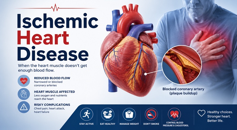
Ischemia means inadequate blood supply to a tissue or organ. When the heart muscle does not receive enough oxygen-rich blood to meet its needs, the result is myocardial ischemia — and the condition that causes this chronically and progressively is ischemic heart disease (IHD).
IHD is also known as coronary artery disease (CAD) and coronary heart disease (CHD). These terms are used interchangeably in clinical practice, and all refer to the same fundamental problem: narrowing or blockage of the coronary arteries, the vessels that supply the heart with blood.
The spectrum of ischemic heart disease runs from silent ischemia — where the heart is oxygen-deprived but the person feels nothing — all the way to sudden cardiac death, with stable angina, unstable angina, and heart attack occupying the middle of the range. What position a person occupies on that spectrum at any given moment depends on how much coronary artery narrowing exists, how abruptly it worsened, and how well the heart is compensating.
How Ischemic Heart Disease Develops: The Role of Atherosclerosis
Nearly all ischemic heart disease begins with atherosclerosis — the gradual buildup of fatty deposits called plaques inside the walls of the coronary arteries.
The process starts silently, often decades before any symptoms appear. Damage to the inner lining of an artery — triggered by high blood pressure, high LDL cholesterol, cigarette smoke, high blood sugar, or inflammation — creates a site where circulating lipoproteins can penetrate the arterial wall. The immune system responds by sending white blood cells to the site. Over years, this accumulation of lipids, immune cells, calcium, and connective tissue forms a plaque that progressively narrows the arterial lumen.
A narrowed coronary artery can still supply enough blood at rest. But during physical exertion or emotional stress, when the heart needs more oxygen and beats faster, the narrowed artery cannot increase blood flow to meet demand. This mismatch between supply and demand is ischemia, and it typically produces the chest pain known as angina.
The more dangerous development is plaque rupture or erosion. Some plaques are structurally unstable — their fibrous cap is thin and their lipid core is large. When such a plaque ruptures, the lipid contents spill into the bloodstream and trigger rapid clot formation at the site. That clot can partially or completely block the artery within minutes, causing the sudden, severe ischemia of an acute coronary syndrome — unstable angina, a heart attack, or sudden death.
The Clinical Spectrum: From Stable Disease to Emergency
Ischemic heart disease does not present the same way in every person. The 2025 American College of Cardiology and American Heart Association guidelines recognize a spectrum of clinical presentations that require different levels of urgency.
Stable Ischemic Heart Disease (Stable Angina)
Stable angina is the most common presentation of IHD, estimated to affect approximately 9.4 million adults in the United States. It is characterized by predictable chest pain or pressure that occurs with a consistent level of exertion and resolves within a few minutes of rest or with nitroglycerin.
The discomfort is typically described as tightening, squeezing, pressure, or heaviness in the center of the chest. It often radiates to the left arm, jaw, neck, or back. It is brought on by walking uphill, climbing stairs, cold weather, emotional stress, or eating a large meal — situations that increase the heart’s oxygen demand.
What makes stable angina “stable” is its predictability. The same activities produce the same symptoms at the same threshold, because the underlying plaque is not changing rapidly. This predictability is clinically important because it signals that the disease is serious but not immediately life-threatening in that moment.
Unstable Angina
Unstable angina is defined by myocardial ischemia that occurs at rest, with minimal exertion, or in a pattern that is new or rapidly worsening compared to previous episodes. It does not fit the predictable pattern of stable angina and signals that something has changed in the coronary artery — typically a plaque that has become more vulnerable or has begun to rupture.
Per the 2025 ACC/AHA guidelines on acute coronary syndromes, unstable angina involves transient myocardial ischemia without significant myonecrosis detectable by cardiac troponin testing. The distinction between unstable angina and a heart attack (myocardial infarction) depends on whether heart muscle cells have actually died — which is detected by troponin elevation in the bloodstream. It is worth noting that widespread use of high-sensitivity troponin testing has changed this boundary significantly: many patients formerly classified as having unstable angina are now reclassified as NSTEMI because modern assays detect even small amounts of myocardial damage.
Unstable angina is a medical emergency. It requires immediate evaluation, not a wait-and-see approach.
Myocardial Infarction: STEMI and NSTEMI
A myocardial infarction — a heart attack — occurs when a coronary artery is blocked severely enough and long enough that heart muscle cells begin to die. There are two major types, distinguished by the pattern seen on an electrocardiogram.
A STEMI (ST-elevation myocardial infarction) results from complete occlusion of a coronary artery, producing characteristic changes on the ECG and causing a large area of heart muscle to be cut off from blood supply entirely. STEMI is the most immediately life-threatening form of heart attack and requires the fastest possible treatment — the goal is to restore blood flow within 90 minutes of hospital arrival.
A NSTEMI (non-ST-elevation myocardial infarction) involves partial rather than complete occlusion, typically producing elevated troponin levels in the blood with different or absent ST-elevation changes on the ECG. While less immediately catastrophic than STEMI, NSTEMI still causes myocardial damage and requires urgent treatment.
Per the 2025 ACC/AHA Acute Coronary Syndromes guidelines, acute coronary syndromes are typically caused by the disruption — rupture or erosion — of an unstable coronary plaque with associated partial or complete thrombosis, resulting in diminished blood flow to the myocardium.
Silent Ischemia
Not all ischemia is felt. A significant proportion of ischemic episodes produce no symptoms whatsoever — no chest pain, no pressure, no shortness of breath. The heart muscle is oxygen-deprived, but the person is unaware. Silent ischemia is particularly common in people with diabetes, elderly patients, and women, whose pain perception may differ from the classic presentation.
Who Gets Ischemic Heart Disease: Risk Factors
Risk factors for IHD are divided into those that cannot be changed and those that can. Understanding them is directly actionable.
Non-modifiable risk factors: Age is the most powerful risk factor. The risk of coronary artery disease rises steeply with age, accelerating after 45 in men and after 55 in women. Male sex confers higher risk earlier in life, though women’s risk rises sharply after menopause and ultimately approaches parity with men. Family history of early heart disease — a first-degree male relative with coronary disease before age 55, or a female relative before age 65 — significantly elevates individual risk.
Modifiable risk factors:
Hypertension damages the arterial wall directly and accelerates atherosclerosis. It is one of the most prevalent and most treatable risk factors for IHD.
High LDL cholesterol provides the raw material for plaque formation. Elevated LDL — particularly in its small, dense form — penetrates arterial walls and initiates the atherosclerotic process.
Tobacco smoking is a potent and underappreciated risk factor. The chemicals in cigarette smoke injure arterial endothelium, promote inflammation, raise blood pressure, lower HDL cholesterol, and increase clotting tendency — attacking nearly every mechanism of cardiovascular protection simultaneously.
Diabetes and insulin resistance substantially accelerate atherosclerosis through multiple mechanisms including endothelial dysfunction, inflammation, abnormal lipid profiles, and elevated blood glucose that directly damages blood vessel walls.
Obesity, particularly abdominal obesity, is associated with insulin resistance, hypertension, dyslipidemia, and systemic inflammation — a cluster of risk factors sometimes called metabolic syndrome.
Physical inactivity independently increases cardiovascular risk through multiple mechanisms, and regular aerobic exercise addresses several other risk factors simultaneously.
Chronic stress and depression are increasingly recognized as genuine cardiovascular risk factors, not merely psychological conditions. Both are associated with elevated inflammatory markers, poorer health behaviors, and directly unfavorable changes in autonomic nervous system tone that affect the heart and blood vessels.
| Risk Factor | Relative Risk Contribution |
|---|---|
| Age over 55 (men) / over 65 (women) | Very high |
| Hypertension | High |
| Smoking | High |
| High LDL cholesterol | High |
| Diabetes | High |
| Family history of early CAD | Moderate to high |
| Obesity | Moderate |
| Physical inactivity | Moderate |
| Chronic stress/depression | Moderate |
Symptoms: What Ischemic Heart Disease Feels Like
The classic symptom of ischemic heart disease is angina — chest pain, pressure, tightness, or discomfort that typically occurs with exertion and resolves with rest. But the clinical reality is considerably more varied, and rigid adherence to the classic description leads to missed diagnoses, particularly in women, people with diabetes, and the elderly.
Classic presentation: Central chest pressure or tightness that radiates to the left arm, jaw, neck, or back. Brought on by exertion, cold, or emotional stress. Relieved by rest or nitroglycerin within minutes. Associated with shortness of breath, sweating, nausea, or a sense of impending doom in acute presentations.
Atypical presentations: Women with IHD more frequently experience shortness of breath, jaw pain, nausea, vomiting, fatigue, and back pain rather than classic chest pressure. People with diabetes often have reduced pain sensitivity due to diabetic neuropathy, making silent ischemia more common. Elderly patients may present primarily with fatigue, shortness of breath, or confusion rather than chest pain.
Shortness of breath on exertion, unexplained fatigue, palpitations, and leg swelling may all represent manifestations of ischemic heart disease, particularly when other risk factors are present.
The most important message: do not dismiss chest-related symptoms because they do not exactly match a textbook description. Any new, unexplained chest discomfort, particularly in the presence of risk factors, warrants medical evaluation.
Diagnosis
Diagnosing ischemic heart disease involves a combination of clinical assessment, blood tests, and imaging studies, with the intensity and sequence of testing guided by the acuity of the presentation.
Clinical history and physical examination remain the starting point. The character, location, radiation, timing, and triggers of chest discomfort inform the clinical probability of IHD before any test is ordered.
Electrocardiogram (ECG) is the fastest and most widely available test. In a STEMI, the ECG shows characteristic ST-segment elevation. In unstable angina and NSTEMI, ST-depression or T-wave changes may be present. In stable ischemic heart disease at rest, the ECG may be entirely normal.
Cardiac troponin is the key blood test for detecting myocardial injury. Troponin proteins are released from heart muscle cells when they are damaged or dying. Elevated troponin in a patient with symptoms of ischemia confirms myocardial infarction. High-sensitivity troponin assays, now standard in most hospitals, can detect very small amounts of myocardial damage with high accuracy.
Stress testing evaluates whether the heart shows signs of ischemia when its oxygen demand is increased. This can be done with treadmill exercise ECG, or with a combination of stress and imaging — stress echocardiography or nuclear stress testing — which shows whether blood flow to specific regions of the heart muscle is impaired under stress conditions.
Coronary CT angiography (CCTA) uses a computed tomography scanner to image the coronary arteries directly, identifying plaques and calculating the degree of narrowing without an invasive catheter. It is increasingly used for risk stratification in stable symptoms.
Coronary angiography — also called cardiac catheterization — is the gold standard for visualizing the coronary arteries. A thin catheter is threaded through the arterial system to the heart, and contrast dye is injected to map the coronary arteries on X-ray imaging. It both diagnoses the extent of disease and, in the same procedure, allows treatment with balloon angioplasty and stenting if significant narrowing is found.
Treatment
Treatment of ischemic heart disease operates at three levels: immediate management of acute events, long-term medical management to prevent progression and future events, and procedural or surgical intervention when needed.
Medications
Antiplatelet therapy — primarily aspirin and, in acute coronary syndromes, a second agent such as clopidogrel, ticagrelor, or prasugrel — reduces the clotting tendency that drives acute coronary events. Dual antiplatelet therapy is standard after a heart attack or stenting procedure.
Statins are the cornerstone of cholesterol management in IHD. They reduce LDL cholesterol, stabilize existing plaques, and have anti-inflammatory effects on the arterial wall. High-intensity statin therapy is recommended for all patients with established ischemic heart disease.
Beta-blockers reduce heart rate and myocardial oxygen demand, reducing the frequency and severity of angina episodes. They also reduce the risk of sudden death after a heart attack.
ACE inhibitors and ARBs reduce blood pressure, protect the heart muscle after a heart attack, and slow the progression of heart failure that can develop as a complication of IHD.
Nitrates relax and dilate blood vessels, improving blood flow to the heart and relieving angina. Short-acting sublingual nitroglycerin is used for acute angina episodes; long-acting nitrates are used for prevention.
Anticoagulants including heparin and its derivatives are used in acute coronary syndromes to prevent clot extension and new clot formation.
Percutaneous Coronary Intervention (PCI)
PCI — commonly referred to as angioplasty and stenting — is the most frequently performed invasive treatment for ischemic heart disease. During the procedure, a balloon catheter is threaded through the arterial system to the blocked coronary artery. The balloon is inflated to compress the plaque and open the artery, and a metal stent is deployed to hold the artery open. Modern drug-eluting stents release medication locally to prevent restenosis.
In STEMI, primary PCI performed within 90 minutes of hospital arrival is the preferred treatment and dramatically reduces mortality compared to thrombolytic therapy alone. In stable ischemic heart disease with significant narrowing causing refractory symptoms despite optimal medical therapy, PCI can relieve angina and improve quality of life.
Coronary Artery Bypass Grafting (CABG)
Bypass surgery creates new pathways for blood to reach the heart muscle by grafting vessels — typically taken from the leg, chest wall, or arm — to route blood around blocked sections of the coronary arteries. CABG is preferred over PCI in patients with multivessel disease, disease of the left main coronary artery, or diabetes with complex coronary anatomy, where the long-term outcomes of surgery are superior to stenting.
Lifestyle and Cardiac Rehabilitation
Medical and procedural treatment of IHD is more effective when combined with sustained lifestyle change. Cardiac rehabilitation — structured programs of exercise, education, and psychological support delivered after a heart attack or cardiac procedure — has been shown in multiple studies to reduce mortality, reduce hospital readmissions, and improve quality of life.
The lifestyle modifications with the strongest evidence for reducing cardiovascular risk in IHD are regular aerobic exercise, smoking cessation, following a heart-healthy dietary pattern, achieving and maintaining a healthy weight, and effective management of blood pressure and blood sugar.
The Gender Gap in Ischemic Heart Disease
One of the most clinically important findings to emerge from recent cardiovascular research is the extent to which ischemic heart disease has been underdiagnosed and undertreated in women. A 2024 narrative review found that women with IHD are frequently underdiagnosed and undertreated despite having similar or greater cardiovascular risk compared to men, contributing to delayed care, suboptimal treatment, and poorer short- and long-term outcomes.
Several factors contribute to this disparity. Women more often present with atypical symptoms that do not match the classic male pattern of crushing chest pressure. Historical clinical trials enrolled predominantly male subjects, and risk algorithms were developed primarily from male data. Women are more likely to have disease of the small coronary vessels — microvascular disease — rather than large-vessel obstruction, and this pattern is less readily detected by standard testing.
Awareness of these disparities is the starting point for closing them. Women with cardiovascular risk factors who experience unexplained shortness of breath, fatigue, or jaw pain — even without classic chest pressure — should have these symptoms evaluated rather than dismissed.
Living With Ischemic Heart Disease
A diagnosis of ischemic heart disease is serious but not a sentence of inevitable decline. Many people with IHD live for decades with well-managed disease and good quality of life. The pillars of living well with IHD are consistent medication adherence, regular medical monitoring, cardiac rehabilitation where appropriate, and the sustained lifestyle changes that support cardiovascular health.
Knowing the warning signs that require immediate attention is essential. Chest pain at rest or chest pain that is more severe, more frequent, or occurring at a lower threshold of activity than usual; new shortness of breath; palpitations or syncope; and any sudden neurological symptoms such as facial drooping, arm weakness, or speech difficulty all warrant immediate emergency evaluation.
For current patients and caregivers seeking evidence-based guidance on IHD management, the American Heart Association maintains comprehensive patient resources at heart.org/coronaryarterydisease.
Frequently Asked Questions
What is the difference between ischemic heart disease and a heart attack?
Ischemic heart disease is the underlying chronic condition — the presence of narrowed coronary arteries from atherosclerosis. A heart attack (myocardial infarction) is an acute event that occurs when a coronary artery becomes suddenly and severely blocked, causing heart muscle cells to die. A heart attack is one of the acute complications of ischemic heart disease, but IHD can exist for years without causing a heart attack.
Can ischemic heart disease be reversed?
The atherosclerotic process cannot be fully reversed, but it can be significantly slowed, stabilized, and in some studies partially regressed with aggressive lipid-lowering therapy, lifestyle change, and blood pressure control. More importantly, the risk of heart attack and death can be substantially reduced with treatment even when the underlying plaque cannot be eliminated.
Is ischemic heart disease the same as coronary artery disease?
Yes. Ischemic heart disease, coronary artery disease, and coronary heart disease are different names for the same condition — reduced blood supply to the heart from atherosclerotic narrowing of the coronary arteries.
How is ischemic heart disease different from heart failure?
They are related but distinct conditions. Ischemic heart disease is the cause. Heart failure — where the heart cannot pump blood efficiently — is a possible consequence. When repeated ischemia or a large heart attack damages enough heart muscle, the heart’s pumping function deteriorates, leading to heart failure. IHD is now the leading cause of heart failure globally.
What does the 2025 AHA/ACC guideline say about blood pressure targets in IHD?
The 2025 AHA/ACC hypertension guidelines maintain a treatment threshold of 130/80 mmHg, emphasizing earlier and more intensive blood pressure control to reduce cardiovascular events, stroke, heart failure, and progression of kidney disease in patients with established cardiovascular disease including IHD.